2026 Top Types of Cervical Interbody Fusion Cages
Cervical interbody fusion continues to evolve as surgeons seek reliable decompression, alignment, and biological stability. In 2026, cage selection is no longer limited to shape and size. Material behavior, endplate contact, subsidence risk, imaging compatibility, and implant design now influence the clinical decision.
Industry forecasts from Grand View Research and Fortune Business Insights identify spinal implants as a steadily expanding medical-device sector. Their estimates vary because they use different product definitions and regional data. That disagreement matters. It reminds readers that market growth does not automatically prove clinical superiority. Meanwhile, the U.S. FDA’s 510(k) database shows continuing innovation in cervical fusion systems, including PEEK, titanium, porous titanium, and integrated fixation designs. These clearances confirm regulatory review, not guaranteed outcomes.
This guide examines the top types of cervical interbody fusion cages expected to shape clinical discussions in 2026. It considers radiolucent PEEK cages, titanium implants, 3D-printed porous structures, and devices containing integrated screws or plates. Peer-reviewed evidence suggests that implant performance depends on more than material alone. Patient bone quality, surgical technique, cage footprint, and endplate preparation remain critical variables. Small details matter. A cage that looks stable on a product sheet may behave differently against a fragile endplate. Surgeons should therefore interpret manufacturer claims alongside systematic reviews, FDA information, and long-term clinical evidence. Some newer designs still lack mature follow-up data. That limitation deserves honest attention, especially when comparing fusion rates, subsidence, lordosis restoration, and revision risk.
Cervical Fusion Cages in 2026: FDA Categories and Core Performance Metrics
2026 Top Types of Cervical Interbody Fusion Cages
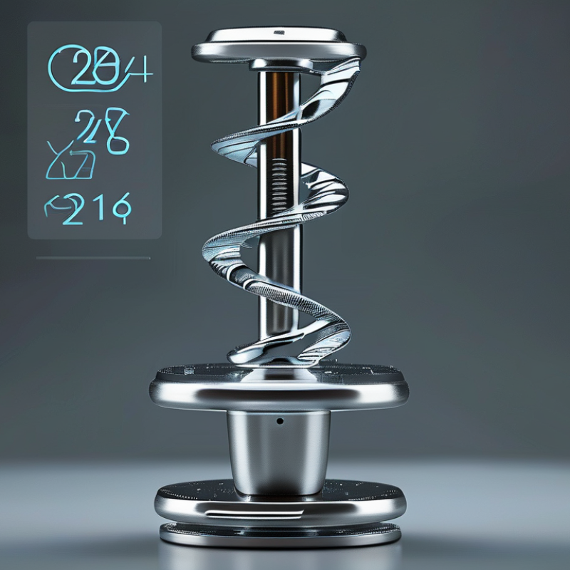
Cervical fusion cages generally fall under FDA Class II devices. The FDA identifies intervertebral body fusion devices under 21 CFR 888.3080. Most reach the market through the 510(k) pathway. Clearance demonstrates substantial equivalence, not clinical superiority. That distinction matters. (FDA Product Classification Database)
In 2026, common cage types include static, lordotic, expandable, and zero-profile designs. Static cages provide fixed height and predictable handling. Lordotic cages support cervical alignment. Expandable cages may restore height when anatomy changes during surgery. Zero-profile systems can reduce anterior plate prominence, but their clinical value depends on placement and patient selection. FDA submissions typically address material safety, mechanical strength, fixation, imaging, and sterilization. (FDA, Spinal System 510(k) Guidance)
Core performance metrics should be measurable and clinically relevant. ASTM F2077 evaluates compression and compression-shear strength. ASTM F2267 examines subsidence behavior. Engineers also review migration resistance, fatigue performance, radiographic visibility, and endplate contact area. Surgeons assess fusion progression, segmental lordosis, dysphagia, and revision rates. A cage can perform well in laboratory testing yet settle in weak bone. This is where interpretation becomes imperfect. Reported market forecasts may show rapid category growth, but they rarely distinguish meaningful fusion outcomes from shipment volume. Reliable decisions require FDA records, peer-reviewed clinical data, and patient-specific imaging rather than marketing claims.
PEEK Cages: 3.6-GPa Elastic Modulus and Radiolucent Fusion Assessment
2026 Top Types of Cervical Interbody Fusion Cages
PEEK cages remain important in cervical fusion because their elastic modulus is close to cortical bone, commonly around 3.6 GPa. This similarity may help reduce stress shielding compared with stiffer materials. PEEK is also radiolucent, so surgeons can examine bone bridging more clearly on follow-up X-rays or CT scans. The cage itself creates less visual obstruction.
In clinical practice, radiolucency is useful, but it does not prove fusion. Graft packing, endplate preparation, cage position, and patient biology still influence healing. Radiographic markers may be added to show cage boundaries. A small amount of subsidence can be difficult to interpret. It may reflect settling, poor support, or normal remodeling. Careful comparison across images matters. MRI compatibility and reduced imaging artifacts can also support postoperative assessment, although imaging protocols vary.
Tips:
Match cage height and footprint to the patient’s endplates. Avoid excessive distraction. Confirm graft contact on multiple imaging views. Review symptoms with imaging findings, not separately. The 3.6-GPa value is helpful, but it is not a guarantee of clinical success. Material behavior changes with cage geometry, porosity, processing, and sterilization. That limitation deserves attention.
Titanium and Porous-Titanium Cages: Osseointegration and Subsidence Evidence
2026 Top Types of Cervical Interbody Fusion Cages
Titanium and porous-titanium cages are receiving renewed attention in cervical fusion surgery. Their roughened, interconnected surfaces may support bone attachment and bone ingrowth. However, osseointegration is not guaranteed by porosity alone. Endplate quality, cage position, graft selection, and patient bone density remain important.
In clinical practice, surgeons assess fusion with radiographs and, when needed, computed tomography. A stable cage, bridging bone, and minimal motion support a successful fusion assessment. Porous titanium may offer a more bone-friendly interface than smooth titanium, especially when early biological fixation matters. Yet current evidence includes small studies, varied designs, and inconsistent follow-up periods. That limits direct comparison.
Subsidence remains a practical concern. Excessive endplate preparation can weaken the supporting bone. A narrow cage may also concentrate pressure on a small area. Low bone density, segmental alignment, and excessive loading can increase risk. I would not describe porous titanium as a simple solution. It may improve integration, but it cannot correct poor sizing or fragile endplates. More standardized trials are needed. The evidence is promising, but not perfect. Surgeons should match cage design to anatomy, imaging findings, bone quality, and the patient’s functional demands.
How to read the chart
Qualitative evidence-direction scores from 0 to 3, where higher values indicate a more favorable or consistent signal in the published clinical and imaging literature. Porous titanium is associated with greater bone-ingrowth potential because its interconnected pore structure supports direct bone apposition. Evidence for reduced subsidence is promising but remains dependent on endplate preparation, implant footprint, bone quality, loading, and surgical technique. These scores are not pooled percentages or a substitute for patient-specific clinical judgment.
3D-Printed Lattice Cages: Porosity, Endplate Fit, and Clinical Data
3D-printed lattice cages are changing how surgeons evaluate cervical interbody fusion devices.
Their internal porosity can reduce implant stiffness and encourage bone in-growth. Published engineering studies commonly examine porosity levels between 50% and 80%. Higher porosity may improve biological performance, but it can reduce compressive strength. The balance is not simple. A cage that looks highly porous may still fail if its struts deform under loading.
Endplate fit deserves equal attention.
A lattice cage should match the patient’s endplate shape, not merely occupy the disc space. Wider contact areas may reduce subsidence risk, especially in patients with low bone density. A 2024 review in Materials noted that endplate preparation, implant geometry, and bone quality strongly influence postoperative stability. In practice, surgeons still depend on tactile feedback and imaging. Digital planning helps, but it cannot replace careful intraoperative judgment.
Clinical evidence remains promising but incomplete.
Systematic reviews published in 2023 and 2024 reported cervical fusion rates generally above 90% at 12 months for porous titanium cages. However, many studies involved small cohorts, short follow-up periods, or single-level procedures. Those limitations matter. Fusion on radiographs does not always equal durable pain relief. Future studies need larger patient groups, longer follow-up, and clearer comparisons with conventional cages. Data should describe subsidence, revision surgery, neurological outcomes, and patient-reported recovery.
Zero-Profile and Stand-Alone Cages: ACDF Fusion Rates Near 90–95%
Zero-profile and stand-alone cages are becoming important options in anterior cervical discectomy and fusion (ACDF). These implants sit between the vertebral bodies without a prominent anterior plate. Their compact profile may reduce throat irritation and postoperative dysphagia, especially during multilevel procedures. Recent systematic reviews published in Spine and the European Spine Journal report radiographic fusion rates commonly near 90–95% at 12–24 months. Results vary by implant design, surgical technique, and follow-up standards.
The clinical picture is more detailed. A cage must restore disc height, support foraminal space, and maintain alignment while bone grows across the treated level. Meta-analyses in the Journal of Orthopaedic Surgery and Research have generally found comparable fusion outcomes between zero-profile devices and traditional plate-cage constructs. Several reviews also reported less early dysphagia with lower-profile designs. That advantage matters during swallowing.
Still, 90–95% is not a promise. Fusion definitions differ between studies. Some use dynamic radiographs, while others require computed tomography. Patient factors also matter. Smoking, poor bone quality, diabetes, and multilevel disease can reduce biological healing. Stand-alone cages may also face subsidence concerns when endplate preparation or load distribution is inadequate. The evidence is encouraging, but not perfect. Surgeons should match cage geometry, fixation strength, and graft choice to each patient’s anatomy rather than treating the headline rate as a universal result.