In the evolving field of spinal surgery, posterior lumbar interbody fusion (PLIF) stands out as a critical technique. Dr. John Smith, a leading expert in spinal surgery, once stated, “The success of a posterior lumbar interbody procedure hinges on precision and technique.” His insights shed light on the importance of employing the best techniques in PLIF, given its complexities and challenges.
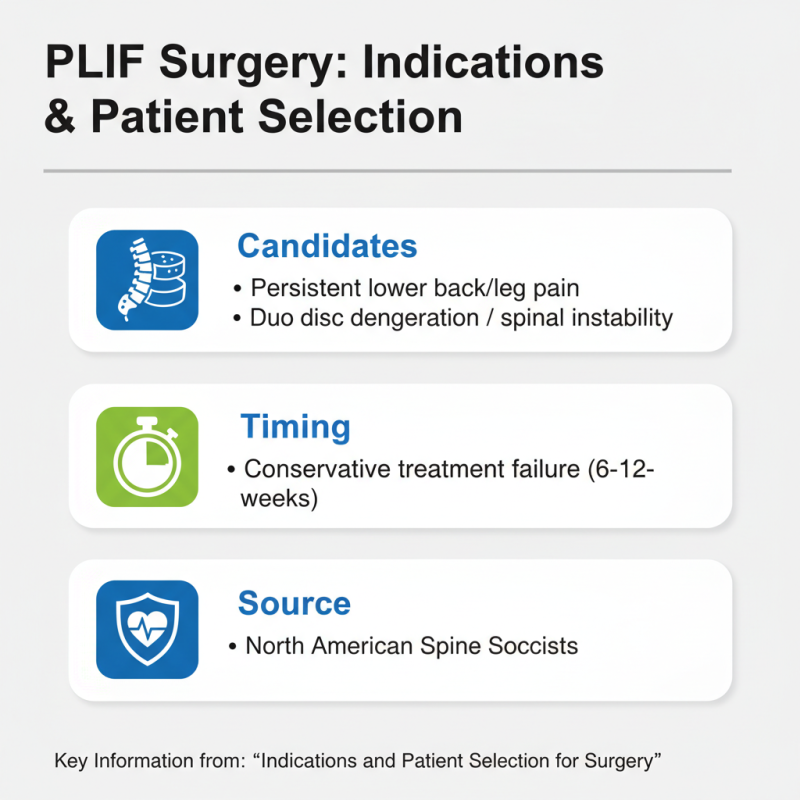
PLIF involves the fusion of the lumbar spine through the back, requiring detailed anatomical knowledge. Surgeons need to be adept at selecting the right approach and instruments. Each technique corresponds to specific indications and patient anatomy. For instance, choosing the right interbody spacer is essential. It can dictate the outcome of the fusion process.
While many techniques are available, none are without risks. Complications can arise, and careful consideration is crucial. Surgeons must reflect on their experiences to enhance future performance. The journey toward mastering posterior lumbar interbody fusion demands continuous learning and adaptation. This emphasis on improvement ensures that techniques evolve, ultimately benefiting patient outcomes.
Posterior Lumbar Interbody Fusion (PLIF) is a surgical method used to treat lumbar spine issues. It involves removing a damaged disc and placing a bone graft between the vertebrae. Surgeons often use instrumentation to stabilize the spine during healing.
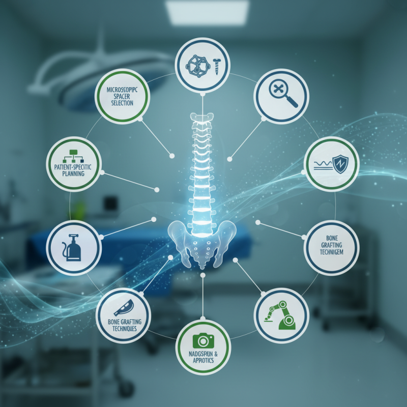
Different techniques exist in PLIF. One method involves a midline approach, allowing access to the spine through the back. This technique can lead to less muscle damage. Another approach focuses on using lateral access. This may reduce recovery time, but it requires specialized training. Both methods showcase the importance of tailored strategies based on patient needs.
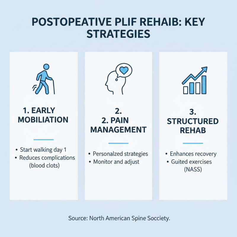
Despite their benefits, challenges arise. Complications can occur, and understanding risks is crucial. Choosing the right technique often depends on the surgeon’s experience and the patient’s unique conditions. Continuous learning in this field is essential, as advancements emerge regularly. Surgeons must evaluate outcomes to improve future practices and enhance patient care.
When considering posterior lumbar interbody fusion (PLIF), patient selection is crucial. Ideal candidates typically exhibit persistent lower back pain or leg pain due to conditions such as disc degeneration or spinal instability. According to the North American Spine Society, surgical intervention is often indicated when conservative treatments fail after 6 to 12 weeks.
Age, overall health, and the presence of comorbidities are critical factors. Patients should ideally be under 75 years old and free of significant cardiovascular or metabolic diseases. A 2020 study revealed that surgery can improve outcomes in up to 80% of carefully selected patients, particularly those with clear structural issues.
Tip: Engage in thorough preoperative assessments. This may include MRI scans to visualize disc issues and spine alignment. Assessments help tailor surgical techniques to individual needs, enhancing recovery potential.
Unexpected outcomes can arise. Some patients may not achieve complete pain relief post-surgery. This emphasizes the need for open discussions about realistic expectations.
Tip: Encourage patients to commit to postoperative physical therapy. This plays a critical role in rehabilitation success, helping patients regain mobility and strength.
Understanding these factors can significantly increase the success rate of PLIF and improve the patient’s quality of life. It’s essential to evaluate each case carefully and continue exploring advancements in surgical techniques.
The posterior lumbar interbody fusion (PLIF) is a complex surgical technique used to stabilize the spine. Surgeons begin by making a midline incision on the patient's back. This incision allows easy access to the spinal column. The muscles and soft tissues are carefully retracted, minimizing damage. Once exposed, the surgeon removes the damaged or diseased disc between the vertebrae. This step is crucial for the success of the procedure.
After the disc removal, the next phase involves placing a bone graft or interbody spacer between the vertebrae. This graft encourages bone growth and fusion. Surgeons may use a variety of techniques to secure the graft in place. Some opt for screws and rods to enhance stability. These come with risks, including potential misalignment. Surgeons must assess their techniques carefully.
The procedure often includes fluoroscopic guidance. This imaging technique helps in visualizing the surgical site in real-time. It may reduce complications but not eliminate them. Healing time can vary, with some patients experiencing discomfort for each stage of recovery. Continuous follow-up with medical professionals is vital for tracking progress. Each case is unique, and reflection on outcomes is essential for improving surgical practices.
Postoperative care after posterior lumbar interbody fusion (PLIF) is crucial for effective recovery. According to the North American Spine Society, implementing a structured rehabilitation program can significantly enhance patient outcomes. Early mobilization is essential. Patients should start walking within a day post-surgery. This practice helps reduce complications such as blood clots and improves circulation. Monitoring pain levels is vital. Effective pain management strategies should be personalized, as each patient responds differently to postoperative pain.
Physical therapy plays a key role in recovery. Engaging in targeted exercises can help restore strength and flexibility. Studies show that patients who participate in physical therapy early tend to regain mobility faster. However, not all patients adhere to rehabilitation protocols. This noncompliance can lead to setbacks. Patients often struggle with pain and fatigue, making it challenging to follow through. Regular follow-ups with healthcare providers can encourage adherence and address any concerns.
A balanced diet is equally important during recovery. Nutritional support can aid in healing and overall well-being. Research indicates that patients with better nutrition heal faster. Hydration also plays a role in recovery, as it supports various bodily functions. While promoting these strategies, it is essential to consider individual patient circumstances. Not every technique will yield the same results for all patients, underscoring the need for tailored approaches in postoperative care and rehabilitation after PLIF.
Surgery for posterior lumbar interbody fusion offers many benefits, but potential complications can arise. One common issue is infection, which may develop at the surgical site. Early signs include increased pain, redness, and fever. Management involves prompt antibiotics and, if needed, further surgical intervention.
Another risk involves nerve damage. This may lead to loss of sensation or motor function. Careful surgical planning and imaging are crucial to prevent this. Surgeons must communicate with patients about the signs of complications, empowering them to seek help early.
Moreover, excessive bleeding can occur during surgery. Surgeons should monitor blood loss and manage it tactically. Understanding the anatomy and using precise techniques can minimize these risks. A comprehensive approach to risk management enhances outcomes. Continual learning and reflection on past surgeries help in improving techniques and reducing complications.
| Technique | Potential Complications | Risk Management Strategies |
|---|---|---|
| 1. Transforaminal Lumbar Interbody Fusion (TLIF) | Nerve injury, blood loss | Intraoperative monitoring, meticulous dissection |
| 2. Anterior Lumbar Interbody Fusion (ALIF) | Vascular injury, retrograde ejaculation | Preoperative imaging, vascular control |
| 3. Posterior Lumbar Interbody Fusion (PLIF) | Postoperative infection, graft failure | Antibiotic prophylaxis, careful graft placement |
| 4. Lateral Lumbar Interbody Fusion (LLIF) | Pneumothorax, thigh paresthesia | Fluoroscopy, patient positioning |
| 5. Interbody Cage Stabilization | Implant migration, subsidence | Proper implant selection, adequate bone preparation |
| 6. Dual Energy X-ray Absorptiometry (DEXA)-guided surgery | Inaccurate bone density measurements | Calibration of equipment, multiple assessments |
| 7. Minimally Invasive Surgery (MIS) | Increased operative time, limited visibility | Advanced imaging techniques, skilled surgical team |
| 8. Bone Morphogenetic Proteins (BMPs) use | Cost, complications from overexpression | Dose optimization, close monitoring |
| 9. Rod and Screw fixation | Hardware failure, neurological complications | Regular follow-ups, using reliable systems |
| 10. Continuous Passive Motion (CPM) | Joint stiffness, discomfort | Patient education, gradual increase in movement |