How to Choose Lumbar Interbody Fusion Implants in 2026?

Choosing a lumbar interbody fusion implant in 2026 requires more than comparing alloy, cage height, or promotional claims. The decision begins with anatomy, instability, bone quality, surgical approach, and the patient’s functional goals. A cage that looks ideal on a product sheet may be unsuitable for a narrow endplate or severe osteoporosis. Small details matter: endplate preparation, graft contact, subsidence risk, and postoperative alignment.

Market reports show strong expansion, but their numbers require careful interpretation. Grand View Research estimated the global spinal implants market at more than USD 12 billion in 2023, while Fortune Business Insights projected continued growth through 2032. These reports often combine cervical, thoracic, lumbar, fixation, and biologic products. Therefore, their figures cannot represent lumbar interbody fusion implants alone. That limitation deserves attention.

Clinical judgment remains central. Spine surgeon Steven R. Garfin, MD, has stated, “The goal of spine surgery is to improve the patient’s quality of life.” The implant should serve that goal, not replace it. Surgeons should review peer-reviewed outcomes, regulatory documentation, subsidence data, fusion rates, revision rates, and long-term follow-up. Real-world experience also matters, especially when comparing expandable, porous, lordotic, and traditional cages.

No implant is perfect. A taller cage may restore foraminal height but increase endplate stress. A porous surface may support bone integration, yet evidence can vary by design and patient group. This guide examines how to balance these trade-offs, question marketing language, and choose lumbar interbody fusion technology with disciplined, patient-specific reasoning.
How to Choose Lumbar Interbody Fusion Implants in 2026?

What Are Lumbar Interbody Fusion Implants?

How to Choose Lumbar Interbody Fusion Implants in 2026?

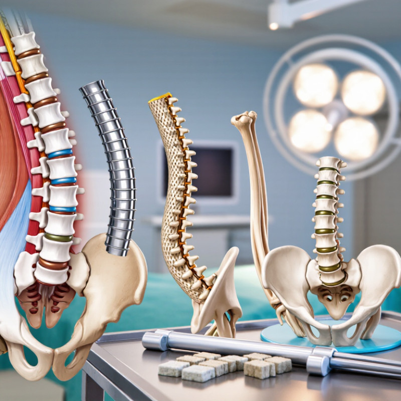
Lumbar interbody fusion implants are medical spacers placed between damaged vertebrae. They replace part of a worn disc and help restore disc height. This can reduce nerve pressure and support bone growth between adjacent vertebrae. Common designs include cages with different shapes, heights, angles, and surface textures. Materials may include titanium alloys, polymers, or combinations of both.

Choosing an implant requires more than viewing a product catalogue. A spine specialist reviews X-rays, MRI scans, bone quality, spinal alignment, symptoms, and previous operations. The implant must fit the patient’s anatomy and surgical approach. It should also provide stable contact with the vertebral endplates. No implant is universally best. Even experienced teams can misjudge size when bone quality is poor. That risk deserves honest discussion.

Tips: Ask how the implant restores alignment, supports bone fusion, and fits your anatomy. Confirm whether bone density testing is needed. Discuss implant height, angle, material, and graft options with your surgeon. Ask what happens if the chosen size does not fit during surgery. In 2026, newer surface treatments and navigation tools may improve placement, but technology does not replace clinical judgment. Good decisions still depend on careful imaging, surgical experience, and realistic recovery expectations. Experimental claims should be questioned.期特码

How to Choose Lumbar Interbody Fusion Implants in 2026?

A lumbar interbody fusion implant, also called an interbody cage, is placed between two vertebral bodies to restore disc height, support alignment, and provide a space for bone fusion. Material selection should consider strength, imaging visibility, bone integration, anatomy, and surgical approach.

The chart compares representative Young’s modulus values. PEEK is closer to the stiffness of bone and generally creates less CT artifact, while titanium alloy provides high structural strength and can support bone-contacting or porous surfaces. Actual values vary by formulation, implant design, porosity, and testing method; modulus alone should not determine implant choice.

Which Patient and Spinal Conditions Require Implant Selection?

Implant selection begins with the patient, not the implant catalog. In practice, lumbar interbody fusion may be considered for painful degenerative disc disease, recurrent disc herniation, spinal stenosis, or unstable spondylolisthesis. Each condition creates different mechanical demands. A collapsed disc may need height restoration, while a slipped vertebra may require stronger correction and stability.

Bone quality changes the decision. Patients with osteoporosis, advanced age, diabetes, or long-term steroid use may face higher risks of subsidence or fixation failure. Imaging should include MRI, CT, and standing radiographs when appropriate. Flexion-extension views can reveal motion that a single scan misses. Previous fusion, scar tissue, spinal deformity, and the number of treated levels also influence implant geometry and surgical approach.

Small details matter. A narrow endplate can limit implant size. Poor sagittal alignment may require more than simple disc replacement with a cage. It is also important to assess frailty, nutrition, smoking status, and the patient’s ability to follow rehabilitation plans. These factors are sometimes underestimated. No implant is ideal for every anatomy. Even careful planning has uncertainty, so surgeons should compare imaging, bone health, symptoms, and long-term goals before choosing a design.

How Do Implant Materials and Designs Affect Fusion Outcomes?

How to Choose Lumbar Interbody Fusion Implants in 2026?

Implant material directly influences bone growth and imaging quality. Radiolucent polymer cages make postoperative CT assessment clearer, while titanium surfaces encourage bone attachment through controlled roughness and porosity. A 2023 systematic review in World Neurosurgery reported one-year fusion rates commonly exceeding 90% with porous or surface-treated titanium designs. Conventional polymer cages often showed lower, more variable rates. Definitions differed across studies. That matters.

Design changes mechanical behavior, not just appearance. Wider footprints distribute load across the endplate and may reduce subsidence. However, aggressive expansion can damage fragile bone. Lordotic cages may improve segmental alignment, yet excessive correction can increase stress at adjacent levels. NASS 2024 guidance continues to emphasize patient selection, endplate preparation, and radiographic follow-up rather than relying on implant geometry alone.

In practice, I would compare material, footprint, height, and surface structure together. A cage that looks biologically advanced may still fail when the endplate is over-prepared. Early clinical reports are encouraging, but long-term comparative evidence remains limited. That gap deserves attention. Surgeons should review bone density, sagittal alignment, smoking status, and fusion goals before selecting an implant. The most expensive design is not automatically the most reliable.

How to Choose Lumbar Interbody Fusion Implants in 2026? – How Do Implant Materials and Designs Affect Fusion Outcomes?

Implant material or design Relevant material/design characteristics Potential effect on fusion and alignment Important limitations or risks Selection considerations
Conventional PEEK cage Radiolucent polymer with an elastic modulus closer to cortical bone than solid metal; allows postoperative assessment of the fusion mass. Produces limited imaging artifact and can facilitate evaluation of bridging bone. Fusion depends heavily on graft quality, endplate preparation, implant stability, and patient biology because untreated PEEK is relatively bioinert. Lower surface bioactivity than porous titanium or bone-derived materials; inadequate endplate support may contribute to settling or subsidence. Useful when radiographic visualization is a priority. Consider surface-treated or textured versions when additional bone–implant interaction is desired.
Textured or surface-treated PEEK Maintains PEEK’s radiolucency while adding a roughened, coated, or otherwise modified surface intended to improve cellular attachment and bone contact. May improve early bone apposition compared with untreated PEEK. Clinical benefit depends on the specific surface technology and available evidence. Surface treatments are not interchangeable; long-term comparative clinical data remain less extensive than the broad clinical experience with standard PEEK and titanium. Review independent clinical data for the exact surface treatment rather than assuming that all modified PEEK products perform identically.
Solid titanium or titanium-alloy cage High strength, established biocompatibility, and a surface that supports bone ongrowth; more radiopaque than PEEK. Provides reliable structural support and immediate stability. Titanium’s radiopacity can make direct assessment of the fusion mass more difficult on CT or radiographs. Higher stiffness than bone may increase stress concentration at the endplate in some configurations; imaging artifact can obscure parts of the fusion region. Consider when high structural strength and established fixation characteristics are important, while accounting for endplate quality and imaging requirements.
Porous or lattice-structured titanium Three-dimensional interconnected porosity can reduce effective stiffness and provide a scaffold for bone ingrowth; pore size, porosity, and manufacturing quality vary by design. May improve bone–implant integration and reduce the mismatch between implant stiffness and host bone compared with solid titanium. Current clinical evidence supports potential benefits but is heterogeneous. Still radiopaque; excessive porosity or insufficient structural support may be inappropriate for certain endplates. Long-term comparative outcome data are developing. Assess verified mechanical strength, pore architecture, endplate-contact area, and independent clinical follow-up.
Carbon-fiber-reinforced PEEK Radiolucent composite designed to combine polymer-like imaging characteristics with increased stiffness and structural strength. Can support visualization of the fusion bed while providing greater rigidity than conventional PEEK. Fusion performance remains dependent on graft, fixation, endplate preparation, and patient factors. Less extensive contemporary clinical evidence than the most widely used PEEK and titanium platforms; material behavior and radiographic markers differ among designs. Consider when radiolucency and structural performance are both priorities, provided the specific implant has appropriate regulatory and clinical support.
Large-footprint cage Uses a broader area of the vertebral endplate, ideally distributing load over stronger peripheral regions rather than the weaker central endplate. May reduce subsidence risk and improve segmental support when the implant is correctly positioned and the endplate is preserved. Oversizing, aggressive endplate preparation, or malposition can injure the endplate or create neural, vascular, or psoas-related risks depending on the surgical approach. Match footprint to vertebral anatomy and approach. Do not sacrifice endplate integrity merely to obtain a larger implant.
Lordotic or wedge-shaped cage Provides a built-in angle intended to restore disc height and segmental lordosis. Can improve segmental alignment and foraminal height when selected for the patient’s anatomy; restoration of overall sagittal balance requires consideration of adjacent levels and pelvic parameters. Excessive correction, anterior malposition, or uneven endplate contact may increase endplate loading, subsidence, or neural symptoms. Use patient-specific imaging and alignment goals rather than choosing the largest available angle.
Expandable cage Inserted at a smaller profile and expanded in situ to increase disc height, foraminal height, or segmental lordosis. May help achieve restoration through less invasive access and can be useful when controlled height adjustment is needed. Over-expansion can damage endplates or neural structures. More moving parts may increase cost and mechanical complexity; long-term evidence varies by design. Use controlled expansion with attention to endplate resistance, neurologic status, implant position, and postoperative imaging.
Integrated fixation features May include screws, anchors, teeth, ridges, or anti-migration features incorporated into or used with the cage. Can improve initial resistance to migration and maintain implant position, which supports the mechanical environment required for fusion. Fixation does not compensate for poor endplate preparation, inadequate graft, osteoporosis, or incorrect implant placement. Prominent hardware may cause approach-specific complications. Select according to surgical approach, bone quality, instability pattern, and whether supplemental posterior fixation is planned.
Graft window and graft volume The internal opening allows placement of autograft, allograft, or a bone graft substitute; volume and access vary with cage geometry. Adequate, well-contained graft in contact with viable bone is essential for bridging fusion. A larger window alone does not guarantee union. Insufficient graft packing, graft migration, poor vascularity, smoking, diabetes, osteoporosis, and multilevel surgery may reduce fusion probability. Evaluate graft containment, packing access, endplate contact, and the patient’s modifiable risk factors together.
Overall evidence-based decision factors Fusion is determined by the interaction of implant material, geometry, stability, graft biology, endplate preservation, fixation, alignment, and patient health. No single material or design is universally superior. Successful fusion generally requires a stable construct, vascularized bone environment, adequate graft, and appropriate mechanical loading. Comparisons are complicated by different surgical approaches, supplemental fixation strategies, follow-up periods, definitions of fusion, and patient populations. Prioritize anatomy, bone quality, sagittal alignment, approach-specific safety, imaging needs, and independent clinical evidence for the exact implant configuration.

Note: Fusion outcomes are multifactorial. Material and design characteristics should be evaluated together with patient-specific anatomy, bone quality, surgical approach, graft strategy, supplemental fixation, and risk-factor optimization.

How Should Implant Size, Shape, and Placement Be Determined?

Choosing a lumbar interbody fusion implant in 2026 starts with the patient, not the catalog. Implant size should reflect disc-space height, endplate width, bone quality, and the correction required. A tall implant may restore foraminal height, but excessive distraction can increase facet stress or endplate injury. Small details matter. Preoperative CT can reveal endplate weakness, while MRI clarifies disc collapse, stenosis, and nerve compression. Standing radiographs show alignment under load.

Shape should match the surgical corridor and endplate anatomy. A broad footprint can support stronger peripheral bone and may reduce subsidence risk. It must still fit the corridor safely. A lordotic shape can support segmental alignment, but the planned angle should respect global balance rather than chase a number. Placement is equally important. The implant should sit centrally or follow the planned correction, with broad endplate contact and no posterior migration. Fluoroscopy, navigation, and tactile assessment can help verify depth and orientation. Technology helps.

The final choice involves stability, decompression, alignment, and biological fusion potential. Osteoporosis, smoking, previous surgery, and sagittal imbalance can change the plan. Surgeons should reassess sizing after preparation, because aggressive endplate removal can make an ideal measurement unreliable. This is where judgment remains imperfect. A template may suggest one size, while the prepared endplate tells another story. The safest decision comes from correlating imaging, intraoperative findings, and the patient’s anatomy. The rationale should be documented for that specific level.

What Safety, Regulatory, and Cost Factors Matter in 2026?

Choosing a lumbar interbody fusion implant in 2026 requires more than comparing catalog prices. Safety should guide the decision. Review evidence for subsidence, migration, infection, and neurological complications. Match implant geometry to the patient’s anatomy and surgical approach. Imaging compatibility also matters, especially when future scans may be needed.

Ask whether the device has current authorization in the treatment jurisdiction. Check its intended use, labeling, sterilization records, and traceability information. Regulatory clearance does not guarantee suitability for every patient. It confirms a defined level of review, not a perfect clinical outcome. That distinction is easy to miss. Surgeons should examine peer-reviewed data, post-market reports, and independent clinical experience. Hospital teams also need clear protocols for handling recalls or adverse events.

Cost analysis should include the entire episode of care. Include the implant, instruments, operating-room time, staff training, imaging, rehabilitation, and possible revision surgery. A cheaper device may create higher costs if it increases complications or inventory waste. Conversely, a premium option may not offer meaningful value for every case. I would request a transparent quotation and compare it with documented outcomes. Local reimbursement rules can change quickly in 2026. Financial assumptions need regular review. Even experienced teams can overlook indirect costs. Patient-specific planning, informed consent, and shared discussion remain essential when evidence is incomplete.