2026 Best Cervical Fixation System for Global Buyers
Choosing the 2026 best cervical fixation system requires more than comparing prices or product photographs. Global buyers need evidence, surgical practicality, and dependable supplier support. A system may look advanced, yet fail to suit a hospital’s instruments, training level, or patient population.
This guide examines cervical fixation through the details clinicians and procurement teams can verify. It considers screw design, plate profile, rod or cage compatibility, imaging visibility, and intraoperative handling. Sterilization instructions matter. So do traceability records, quality certifications, clinical documentation, and post-market surveillance. These details often decide whether a system performs smoothly in a real operating room.
Small differences matter.
Experienced surgeons may assess screw angulation with gloved hands, limited visibility, and changing anatomy. Buyers may also compare packaging, delivery reliability, technical training, and local service capacity. Regulatory expectations differ between markets, so international purchasing teams should confirm current requirements with qualified local experts. Manufacturer claims should never replace independent clinical judgment or hospital approval processes.
No single cervical fixation system is best for every case or region. That point deserves emphasis. A familiar system may offer safer workflow, while a newer design may provide useful flexibility but limited long-term evidence. This article presents a practical, evidence-aware framework for comparing leading options in 2026. It also recognizes an uncomfortable reality: product selection can remain uncertain when data are incomplete, marketing language is strong, and real-world results vary.
Cervical Fixation Systems Explained: Anatomy of the 7-Vertebra Cervical Spine
2026 Best Cervical Fixation System for Global Buyers
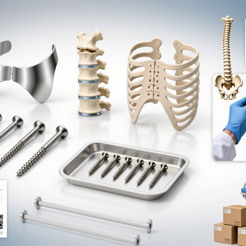
The cervical spine contains seven vertebrae, labeled C1 through C7. Together, they support the head and protect the spinal cord. C1 forms a ring around the upper spinal cord. C2 provides the pivot for head rotation. C3 to C7 gradually become larger and carry more load. C7 often creates the visible bump at the base of the neck.
Between most vertebrae, discs absorb movement and reduce impact. Facet joints guide bending and rotation. Small openings allow nerve roots to exit toward the shoulders, arms, and hands. The vertebral arteries also travel through openings in the upper cervical region. This anatomy makes implant placement highly precise. A fixation system must match bone quality, alignment goals, and the treated levels. Imaging should confirm screw paths, disc spaces, and nearby neural structures before surgery. In practice, small anatomical differences matter. A standard approach may still need careful adjustment.
Tips: Check C1–C7 anatomy on high-quality imaging. Compare implant dimensions with local bone structure. Confirm that instruments support controlled placement. Ask whether the design preserves necessary movement at untreated levels. Do not judge a system by appearance alone. That is an easy mistake. Surgical training, documented testing, and regulatory compliance also deserve review. Product information can be clear, yet clinical judgment remains essential.
2026 System Types: Anterior, Posterior, and 360° Fixation Configurations
For global buyers, cervical fixation should be selected by anatomy, pathology, and surgical access.
Anterior systems support direct decompression and often use plates, screws, or stand-alone devices. Posterior systems provide strong fixation when instability, deformity, or multilevel disease demands broader support. A 360° configuration combines anterior and posterior constructs. It can improve stability, but it also increases surgical exposure and planning demands.
The 2024 AHRQ evidence review reported that outcomes vary with diagnosis, surgical level, and patient risk, rather than implant choice alone. This matters. A 2024 Grand View Research analysis also projected steady cervical spine implant market growth through 2030, with an estimated annual rate near 5% to 6%. Buyers should not treat market growth as proof of clinical superiority. Evidence remains uneven across configurations. That weakness deserves attention.
Tips:
Request radiographic examples, fatigue-testing data, sterilization instructions, and published clinical evidence. Check screw-angle control, locking security, and compatibility with local instruments. Anterior constructs may suit focal decompression, while posterior fixation can better manage long-segment instability. Combined 360° surgery may be reasonable for selected high-risk patterns, but patient condition and surgeon expertise remain decisive. Fit matters more. Not always.
Clinical Benchmarks: Fusion Rates, Revision Risk, and 10–15° Motion Targets
2026 Best Cervical Fixation System for Global Buyers
Clinical purchasing should begin with measurable outcomes, not catalog features. Published FDA-reviewed cervical studies commonly report single-level fusion rates above 90% at two years. However, multilevel procedures show lower consistency, especially when bone quality, smoking, or poor alignment affects healing. A 2023 systematic review in Spine also linked pseudarthrosis with higher reoperation exposure, although reported revision rates varied widely by construct and follow-up period.
Motion targets require equal caution. In motion-preservation studies, maintaining approximately 10–15° of segmental movement is often considered clinically meaningful. Excessive motion is not automatically beneficial. A poorly controlled segment may increase facet loading or create uneven wear. European Spine Journal reviews have shown that preserved movement does not always produce superior pain scores. The number looks attractive. The patient matters more.
For global buyers, supplier evidence should include radiographic fusion definitions, minimum 24-month follow-up, revision causes, and independent adverse-event reporting. Ask for subgroup data from osteoporotic patients and multilevel cases. A low revision rate may reflect short observation. That is a real weakness. Surgical experience also changes outcomes, so laboratory testing cannot replace clinical evidence. The strongest evaluation combines fatigue testing, screw pullout data, validated patient-reported outcomes, and transparent post-market surveillance.
2026 Best Cervical Fixation System for Global Buyers
Clinical Benchmarks: Fusion Rates, Revision Risk, and 10–15° Motion Targets
The chart presents non-brand-specific clinical reference ranges commonly reported for cervical procedures: approximately 90–95% radiographic fusion, 5–10% revision risk over medium-term follow-up, and a 10–15° segmental motion target for motion-preserving cervical constructs. Actual outcomes vary by indication, surgical technique, patient selection, follow-up duration, and definition of revision.
Implant Selection Criteria: Materials, Screw Angles, and MRI Compatibility
Selecting a cervical fixation system in 2026 requires more than comparing catalog prices. In operating rooms, material behavior affects handling, imaging, and long-term follow-up. Titanium alloys remain common because they combine strength with relatively low MRI artifact. PEEK components may reduce imaging distortion, but their load-sharing role must match the construct design. Stainless steel can be strong, yet it may create more artifact and demands careful MRI review. Material names alone are not enough. Verify composition, surface treatment, fatigue data, and sterilization instructions from technical files.
Screw angle is a practical selection issue, not a decorative feature. Variable-angle screws can help surgeons adapt to patient anatomy and plate position. Fixed-angle screws may provide predictable locking in selected constructs. Too much angular freedom can complicate placement, especially near narrow bony corridors. The best range depends on vertebral level, bone quality, and the planned surgical approach. Cadaveric data can inform choices, but it cannot replace clinical judgment. I would question any claim promising one angle for every anatomy.
Tips:
Ask for MRI conditions at 1.5T and 3T, including field strength, SAR limits, scan duration, and artifact measurements. Confirm whether the complete construct, not only one screw, was tested. Review pullout and fatigue results. Request traceable lot information and clear labeling. A small documentation gap can become a large imaging problem. Recheck local regulatory requirements before procurement.
Global Buyer Checklist: FDA, CE, ISO 13485, and Sterilization Compliance
2026 Best Cervical Fixation System for Global Buyers
Global Buyer Checklist: FDA, CE, ISO 13485, and Sterilization Compliance
Selecting a cervical fixation system requires more than reviewing implant design or surgical preferences. Buyers should verify the device’s regulatory pathway in each target market. In the United States, check the applicable FDA clearance or approval, device classification, indications, and manufacturing site details. A registration record alone does not prove market authorization. Review the official documents carefully.
For European distribution, confirm compliance with the Medical Device Regulation and verify the CE certificate’s scope, validity, and notified body information. The certificate should cover the exact system, accessories, and intended use. ISO 13485 certification supports quality management, but it does not replace regulatory approval. Ask for audit status, complaint procedures, corrective action records, and traceability controls. These details often reveal more than a polished product brochure.
Sterilization evidence deserves close attention. For sterile products, request validation records for the stated method, such as ethylene oxide, radiation, or moist heat. Check relevant standards, including ISO 11135, ISO 11137, ISO 17665, and ISO 11607, when applicable. Packaging integrity, shelf-life data, residual limits, and transport testing should match the product’s real distribution conditions. A warehouse exposed to heat and humidity may challenge a perfect laboratory result.
Experienced buyers still miss small gaps. I have seen certificates with unclear product families and outdated addresses. That is not always fraud, but it demands clarification. Request the declaration of conformity, instructions for use, risk-management evidence, and post-market contact details before approving a supplier. Keep every version controlled. Credibility is built through traceable evidence, not confident language.